Imaging science: A view through space and time

Dr. Pavan Vedula’s snapshot of Wistar’s Imaging Facility
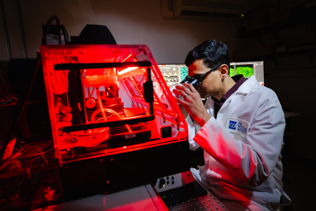
Cytoskeletal biologist Dr. Pavan Vedula joined Wistar’s Imaging Facility in January as managing director. He brings enthusiasm and zeal to his job delivering the most sophisticated imaging methods and equipment. Using the cutting-edge optical instruments he manages, Wistar scientists can analyze cells & tissues in fresh innovative ways and advance a multitude of preclinical studies in cancer and infectious disease.
How do you describe what Wistar’s Imaging Facility does?
Pavan Vedula: Scientific imaging strives to answer questions arising from what we observe; it’s a mechanism to observe and/or visualize biology directly. At Wistar’s Imaging Facility, you can observe the nature of biology (cells & tissue) from a macro level—at the size of large-scale objects, like an organ—to the nanometer level, like two proteins interacting.
We can use imaging to understand biology in the context of space and time down to single cell or single molecule level. Unlike other techniques, where you may be looking at an entire population or the contents of a test tube, with our imaging equipment you can look at specimens live or dead, in their spatial context and over time.
Why is it important to see biology in all these different contexts?
PV: The many ways in which you look at the nature of biology allows you to address different questions. Some questions can only be answered when observed in the spatial and/or temporal context, which is where the Imaging core comes in. In certain other cases, you can utilize the technologies at multiple cores to synergistically answer questions in biology. Lastly, imaging can serve as a complimentary approach to validating results derived through other techniques. These are all useful in terms of asking different questions in science.
How quickly is imaging technology progressing and how does that help us advance science?
PV: Just over the short span of my career, I have seen the field of microscopy grow from looking at slow spatial or temporal context, to the nanometer and microsecond scale. This kind of cutting-edge microscopy is no longer limited to a specialized lab with an optical physicist. Now anyone can, with the click of a few buttons, look at things on the nanometer scale and understand biology and context on that nanometer scale. Cameras are so fast you’re able to see things happening at a much faster timescale. If you imagine looking macroscopically at the organ, then the cell, then proteins; proteins are so small that things are happening quickly at this scale. It’s been a fascinating journey seeing the evolution and democratization of imaging technology that now allows us to capture biology at this level.
Regarding innovation and the future, where do you see the potential for imaging to transform science in the next 10 years?
PV: One aspect of technological innovation is the integration of image analysis and workflow tools. AI driven image analysis allows for automation in data acquisition by quickly analyzing, for instance, smaller scale of data and homing in on exactly where you need to image. These workflows give people time to do more science rather than sitting in front of a microscope for three hours and just staring at the microscope imaging slowly.
Another aspect is being able to generate hypotheses with large data sets that microscopes produce these days, edging into a bioinformatics kind of space. You’re able to have multiplexed images where you have 15-16 or sometimes up to 100s of dimensions that light-up your spatial context like never before. These high-plex images are being used to address complex questions and understand more deeply the science happening in the spatial context.
What’s one thing about microscopic life that still excites you, no matter what you’re doing or how many times you see it?
PV: I have a soft spot for the cytoskeleton because I was a cytoskeletal biologist for 13 years. I love all things cytoskeleton. Every time I see it, I’m like, oh, that’s just gorgeous.