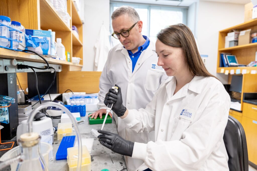
Breakthrough Research at Wistar

Pioneering Advances in Biomedical Science
Wistar researchers continue to push the boundaries of scientific understanding, developing innovative approaches to address complex medical challenges in cancer, immunology, and infectious disease.
Revolutionary Vaccine and Immunotherapy Platforms
Dr. David B. Weiner, Executive Vice President, director of Wistar’s Vaccine & Immunotherapy Center, and W.W. Smith Charitable Trust Distinguished Professor in Cancer Research, and collaborators have achieved a transformative breakthrough in antibody delivery systems. In the first successful human trial of its kind, researchers demonstrated that synthetic DNA can direct human cells to produce functional monoclonal antibodies against COVID-19 that persist in the bloodstream for over a year.
The Phase 1 clinical trial showed that DNA-encoded monoclonal antibodies (DMAbs) were detected in 100% of evaluable participants, with sustained expression observed throughout 72 weeks of followup— addressing one of the critical disadvantages of existing antibody biologics, short half-life. Another shortcoming of existing biologics is resistance, or the development of anti-drug antibodies (ADAs). The DMAbs developed by Weiner and his team produced sustained expression after just 1–4 doses with no detectable ADAs. Furthermore, unlike existing RNA vaccines, DMABs do not require cold storage, thereby expanding accessibility and lowering costs. The implications of DMAbs research extend far beyond vaccinations against COVID-19. This technology could deliver long-acting treatments for cancer, autoimmune diseases, and other conditions requiring frequent hospital visits. Paired with its advantages of requiring fewer doses and being more easily storable in resource-limited settings, DMAbs could transform how we prevent and treat diseases globally.
Novel Cancer and Autoimmune Therapeutics
Dr. Maureen Murphy, Ira Brind Professor and Deputy Director of the Ellen and Ronald Caplan Cancer Center, and her lab recently overturned three decades of scientific thinking about p53, the most important tumor suppressor protein in cancer research. In a study published in Molecular Cell, they reveal for the first time that this critical protein, responsible for halting cell division or initiating cell death, changes its binding sites according to specific cellular signals. The findings challenge the long-held scientific belief that p53 always activated the same set of genes regardless of cellular context or outcome. Instead, researchers discovered that p53 can be directed by the enzyme PADI4 to leave some of its usual binding sites and relocate to genes that generate an immune response to attack tumors.
The discovery emerged from Murphy’s decades-long investigation into genetic variants found in families of African descent who develop cancer at accelerated rates but don’t fit typical hereditary cancer patterns. These families carry partially functional versions of p53—called hypomorphic variants—which are understudied and therefore leave patients without clear medical guidance. Murphy and her team hypothesized that studying these semi-functional variants that are associated with cancer would reveal the key target gene for p53 tumor suppression.
Dr. Luis Montaner, Executive Vice President, Director of the HIV Cure and Viral Diseases Center and Hebert Kean, M.D., Family Professor has identified a novel therapeutic target that represents a new approach to treating solid tumors. His research demonstrates how blocking a specific cleft in cells’ retinoblastoma protein can selectively kill tumor-protecting macrophages in ovarian cancer without affecting the protein’s cancer-suppressing abilities.
This discovery emerged from HIV research, where scientists identified retinoblastoma protein’s role in macrophage survival during infection. Montaner’s findings solve a persistent challenge in cancer immunotherapy: targeting immune cells that tumors hijack to protect themselves without compromising beneficial immune functions needed to fight those tumors. By blocking the specific protein cleft, Montaner and his team successfully depleted tumor-protecting macrophages while leaving cancer-fighting macrophages intact, which resulted in tumor shrinkage in animal models. Next, the team will extend applications of this research beyond ovarian cancer, studying how regulating retinoblastoma protein affects macrophages in acute myeloid leukemia and pancreatic cancer. They will also test the approach in combination with immunotherapy.
Dr. Joseph Salvino, in collaboration with The University of Leeds and the University of Pennsylvania’s Perelman School of Medicine, has discovered a new “molecular glue” that shows potential for treating autoimmune diseases by reducing inflammation. This category of “glue,” termed BLUEs (BRISC molecular glues), works by binding to specific overactive proteins to temporarily deactivate them.
The research targets dysregulation in the BRISC protein complex, which normally helps manage inflammatory signaling. When BRISC is overactive, it worsens inflammation, contributing to autoimmune conditions like lupus. Traditional drugs that block activity in the overactive BRISC complex often fail because they are unable to attach with enough specificity to stop the overactivity and consequently cause unwanted side effects. In preclinical trials—including with blood cells from people with scleroderma, an autoimmune disease characterized by high interferon activity—Salvino’s team found that BLUEs bind to BRISC, keeping the complex locked in an inactive state without destroying it. This mechanism reduces the chance of side effects while effectively reducing interferon signaling. This approach represents one of the first times molecular glues have been used to target an enzyme like BRISC, offering new possibilities for treating autoimmune diseases.
Viral Mechanisms and Therapeutic Targets
Dr. Chengyu Liang, professor and co-leader of Wistar’s Molecular & Cellular Oncogenesis Program, has uncovered a previously unknown mechanism by which viruses reprogram mitochondrial structure to silence immune responses. Her research reveals how the Kaposi’s sarcoma-associated herpesvirus (KSHV) protein vBcl-2 hijacks the enzyme NM23-H2 of an infected cell to change the shape of mitochondria such that they cannot mount an immune response to viral replication.
Liang’s research demonstrates that vBcl-2 triggers mitochondrial fragmentation at critical moments in the KSHV lifecycle, preventing assembly of cells’ immune signaling platform, MAVS, that normally triggers antiviral defenses. This sophisticated strategy allows viruses to complete late-stage assembly and exit from infected cells without being waylaid by two key antiviral proteins—TRIM22 and MxB—which would normally be activated by MAVS and trap virus particles in the cell’s nucleus, preventing their release.
The research team also identified a small-molecule compound, VBNI-1, that disrupts this virus-host interaction. In lab models, VBNI-1 blocked mitochondrial fission and restored immune signaling without toxicity to uninfected cells. This candidate drug offers hope for KSHV treatment, for which there is currently no vaccine or cure, and potentially for treatment of other herpesviruses, as well.
Dr. Italo Tempera, associate professor in the Genome Regulation and Cell Signaling Program, has made two groundbreaking discoveries about Epstein-Barr virus (EBV) mechanisms in cancer development and treatment.
Tempera’s team discovered that a class of FDA-approved cancer drugs called PARP1 inhibitors can effectively combat EBV-driven lymphomas through an entirely novel mechanism. Instead of preventing DNA repair, which is how these drugs work to kill most tumor cells, PARP1 inhibitors help to control which genes are accessible and active in EBV infection. By applying a PARP1 inhibitor that has already been approved to treat breast cancer to a mouse model of EBV-driven lymphoma, the researchers found that the drug stops a specific EBV protein, EBNA2, from activating MYC, a gene that promotes cancer. Disrupting EBNA2, and thereby MYC, achieved an 80% reduction in tumor growth in the mouse models without generating the DNA damage typically associated with PARP inhibitor therapy. This approach, which uses drugs that are already on the market, provides new hope for not only patients with lymphoma, but for patients with other EBV-associated cancers that may rely on this mechanism, as well.
In parallel research, Tempera revealed how the EBV protein EBNA-LP, originally thought to be a “helper” protein without a distinct role of its own, fundamentally rewires the three-dimensional structure of DNA in infected B cells. By interacting with the cellular protein YY1, EBNA-LP changes the arrangement of the genome in these immune cells to unlock regions that would normally be restricted. This process converts mature, differentiated B cells into a more naive, stem-cell-like state, making the infected cells more adaptable and responsive to signals that promote cancerous growth.
EBNA-LP joins two other viral proteins, including the aforementioned EBNA2, in affecting how B-cells’ genomes are folded. The fact that the virus has evolved multiple proteins to target the same cellular process signals that this mechanism is critical for infection success—and reveals a vulnerability that may be exploited by EBV-related cancer treatments.